Histology of FALLOPIAN TUBE MEDizzy
Last updated: May 12, 2019 Revisions: 21 format_list_bulleted Contents add The uterine tubes (or fallopian tubes, oviducts, salpinx) are muscular 'J-shaped' tubes, found in the female reproductive tract. They lie in the upper border of the broad ligament, extending laterally from the uterus, opening into the abdominal cavity, near the ovaries.
Fallopian Tube Histology Labeled
fallopian tube, either of a pair of long narrow ducts located in the human female abdominal cavity that transport male sperm cells to the egg, provide a suitable environment for fertilization, and transport the egg from the ovary, where it is produced, to the central channel (lumen) of the uterus.. Each fallopian tube is 10-13 cm (4-5 inches) long and 0.5-1.2 cm (0.2-0.6 inch) in diameter.

Online histology slides atlas Complete student friendly histology atlas Medic For You
The fallopian tubes are bilateral conduits between the ovaries and the uterus in the female pelvis. They function as channels for oocyte transport and fertilization. Given this role, the fallopian tubes are a common etiology of infertility as well as the target of purposeful surgical sterilization. They can also be sites of ascending infection or neoplasms.
Pathology Outlines Anatomy, histology, embryology & features to report
Fallopian histology slide labeled diagram Let's try to identify all the histological features from the actual slide sample with the help of the fallopian histology slide labeled diagram. Suggested slides for you - The ovary histology slide with a labeled diagram A proliferative and secretory uterus histology slide with the labeled diagrams
+histology+slide.jpg)
Histology Slides Database Fallopian Tube histology Slides
1) the trapping of the egg, 2) a region where fertilization takes place, and 3) a region simply for transport to the uterus. Examine its open end near the ovary, the infundibulum View Image, in slide 240-1 and note the funnel shape and ridges of mucosa that extend as a fringe of finger-like projections known as fimbriae.

Uterine/Fallopian Tube Fallopian tubes, Histology slides, Female reproductive system
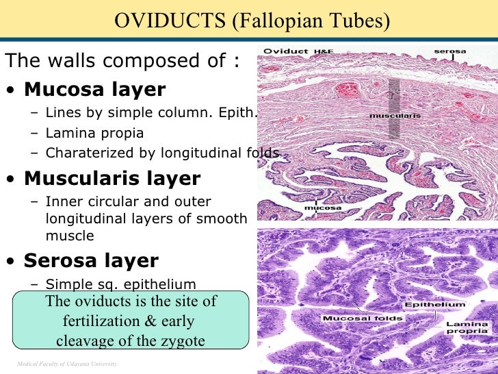
UTERUS UTERINE (FALLOPIAN) TUBES VAGINA PLACENTA MAMMARY GLANDS FEMALE REPRODUCTIVE SYSTEM The female reproductive system consists of 2 ovaries, 2 oviducts (also known as uterine or fallopian tubes), the uterus, vagina, external genitalia and 2 mammary glands.
+histology+slide.jpg)
Histology Slides Database Fallopian Tube histology Slides
The fallopian tube ( TA: tuba uterina 8 ), also known as the uterine tube or, less commonly, the oviduct, is a paired hollow tube that bridges the ovary and uterus and functions to convey the mature ovum from the former to the latter. If conception occurs, it usually does so within the tube, which can be affected by a wide range of pathology.
Fallopian tube Normal Histology NUS Pathweb NUS Pathweb
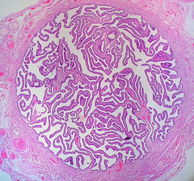
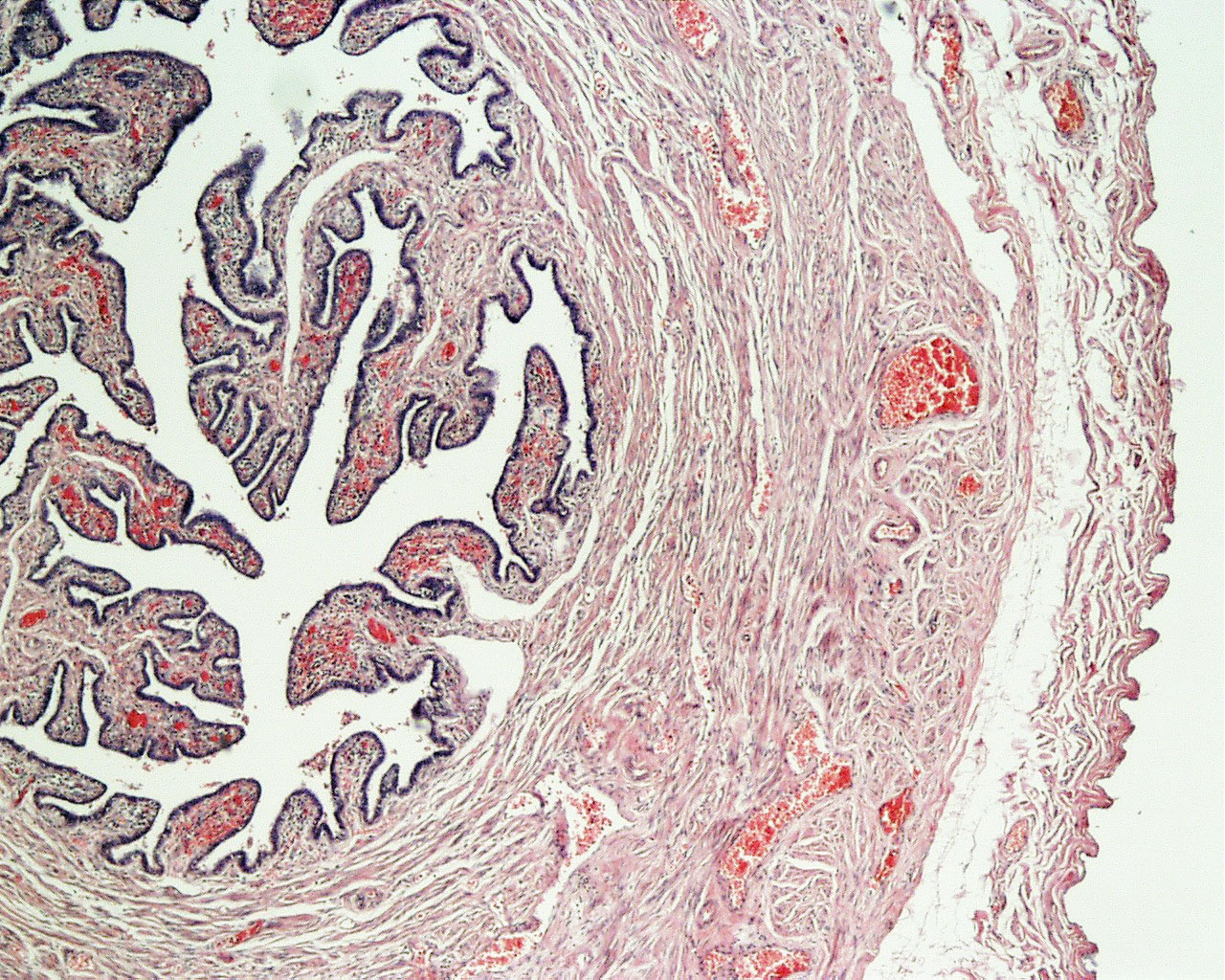
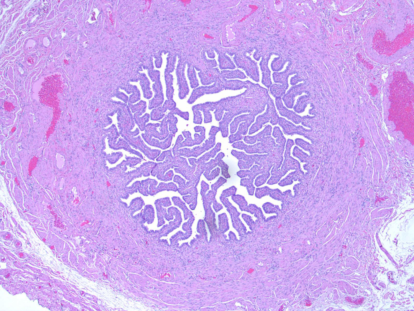
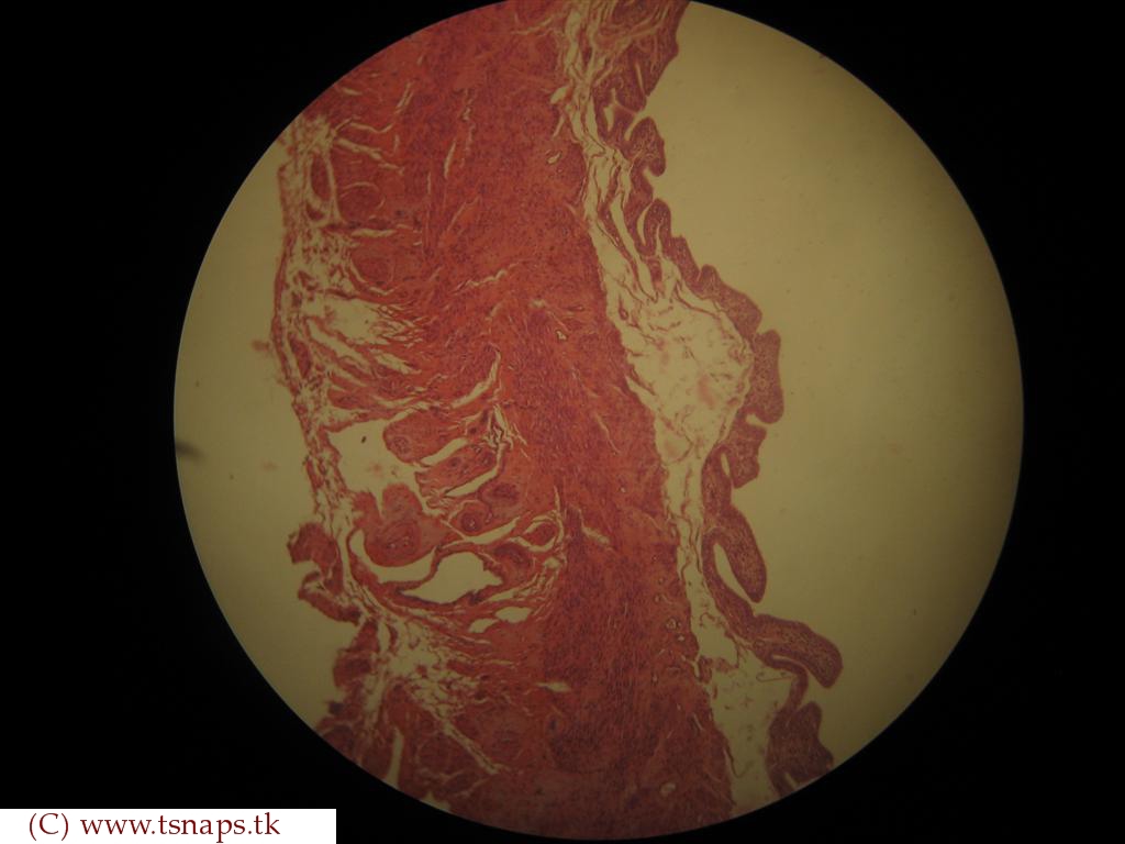
Histology at SIU Fallopian Tube A fallopian tube (also called uterine tube or oviduct) conducts each egg, following ovulation, from the ovary to the uterus. The wall of the fallopian tube includes an elaborately folded mucosa (endosalpinx) surrounded by a muscularis (myosalpinx).
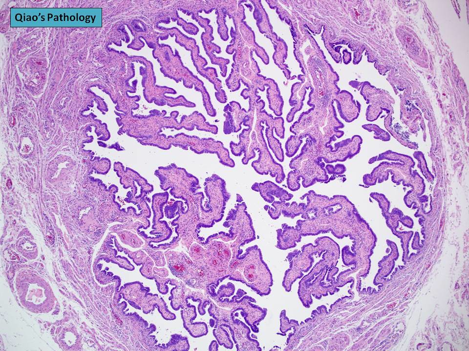
Qiao's Pathology Normal Fallopian Tube (乔氏病理学:正常输卵管) Flickr
Fallopian Tube Chapter Outline Introduction 459 Anatomy, Histology, and Function of the Fallopian Tube 459 Anatomy 459 Histology 460 Mucosal Epithelial Alterations 461 Function 462 Approach to Examining Tubal Specimens 462 Bilateral Tubal Ligation for Sterilization 462 Salpingectomy for Tubal Ectopic Gestation 463
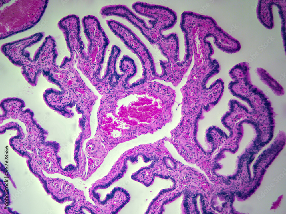
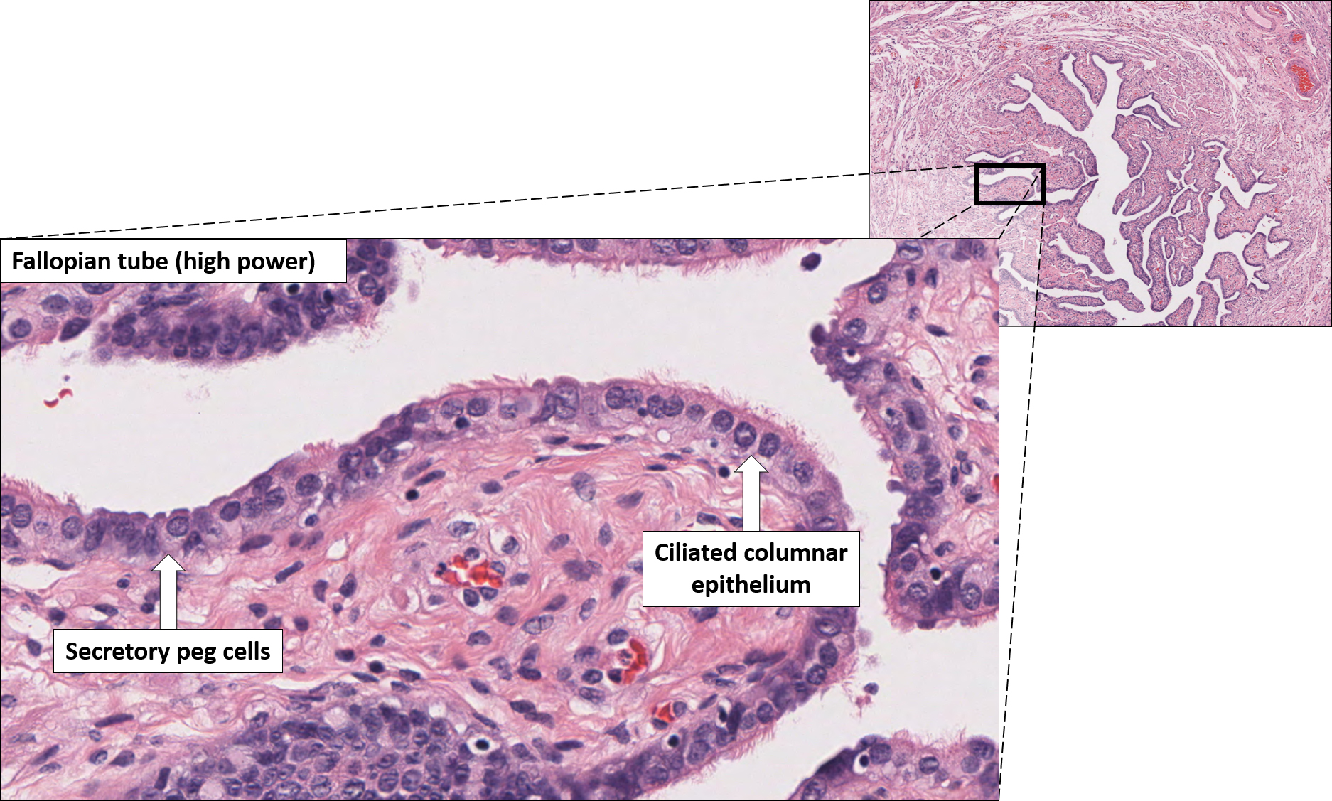
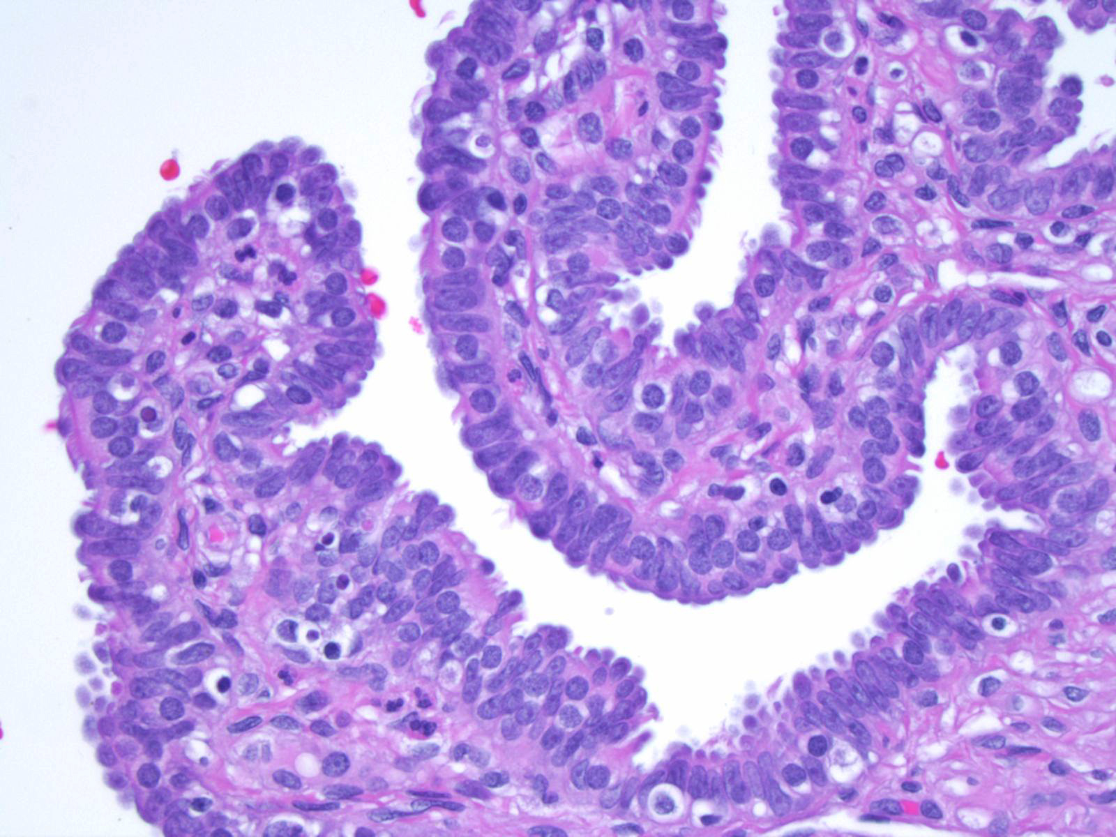
Histology image of human fallopian tube showing ciliated simple columnar epithelia and mucous
Fallopian tube epithelial tumors: carcinosarcoma endometrioid adenocarcinoma high grade serous carcinoma serous adenofibroma and papilloma serous borderline tumor (pending) serous tubal intraepithelial carcinoma Fallopian tube mixed epithelial and mesenchymal tumors: adenosarcoma (pending) Fallopian tube germ cell tumors: teratoma (pending)
Human Biology Online Lab / Copy of Fallopian Tubes by Elynn Johnson
A fallopian tube consists of a thin mucous membrane and layers of muscle. Mucous membrane: A delicate lining in your fallopian tubes secrete fluids that maintain an environment where fertilization can happen and an embryo can develop. Small hair-like structures in the lining (cilia) sway, moving eggs, sperm and an embryo (if fertilization takes.

Active Learning for the Medical Sciences Draw It to Know It
Physiologic functions discussed in this article include the role of the fallopian tube in sperm transport, its part in sperm maintenance and capacitation, and the tube's function in ovum transport, fertilization, and embryo transport. Clinically, the role of the myosalpinx is undetermined, although it may affect tubal motility and ovum transport.
Pathology Outlines Anatomy, histology, embryology & features to report
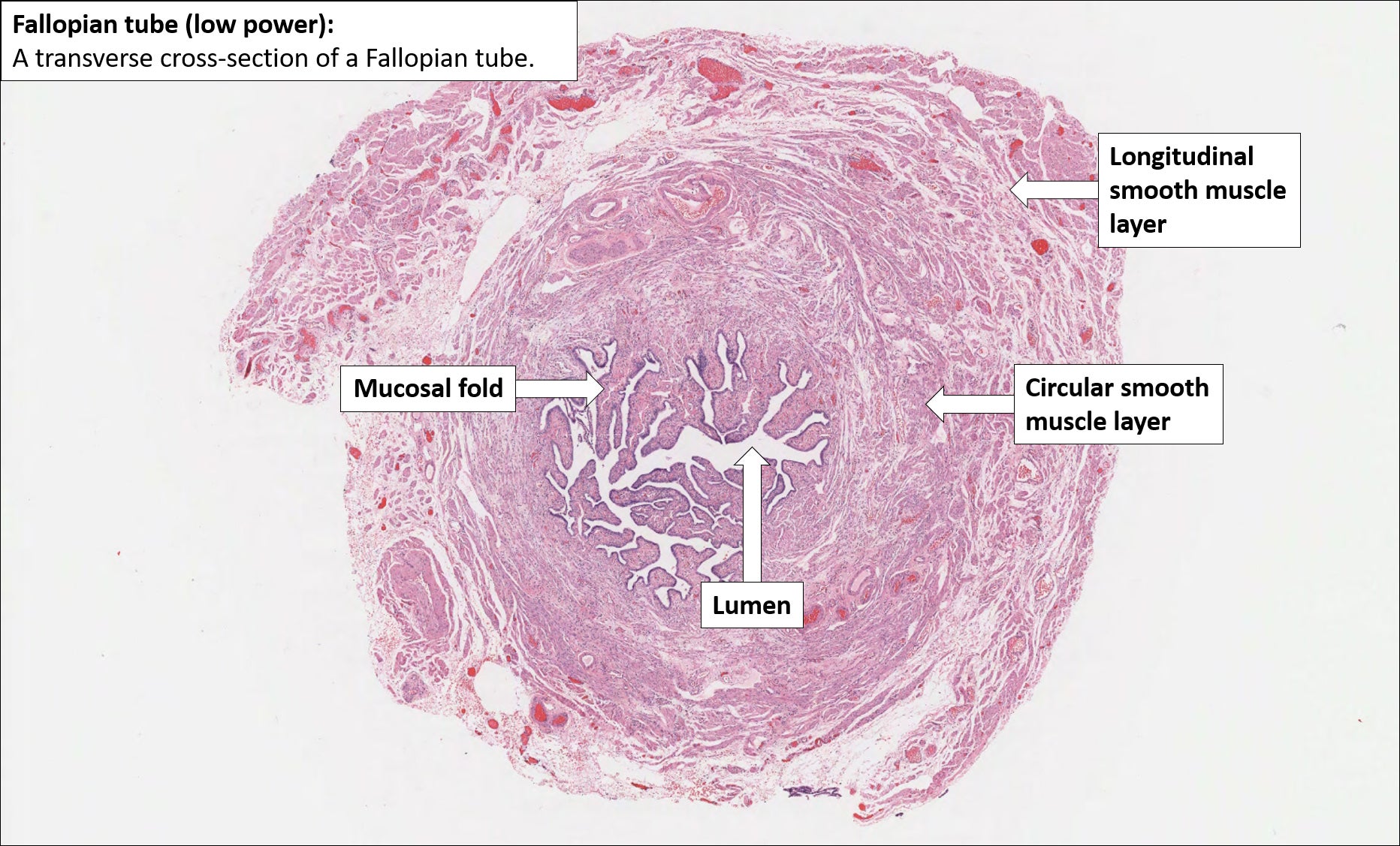
show labels Take a look at this low power image of the fallopian tube. Can you identify the serosa (adventitia layer), muscularis mucosa layer, the mucosa, which is very highly folded longitudinally, and the lumen. The folded mucosa are where the ova are fertilised.
Fallopian tube Normal Histology NUS Pathweb NUS Pathweb
The uterine tubes (a.k.a. fallopian tubes) are important structures in the female reproductive tract, which connect the peritoneal cavity with the uterine cavity. They provide a site for fertilisation and are involved in the transport of the ovum from the ovaries to the body of the uterus. The uterine tubes are also referred to as the oviducts.

Histology of Fallopian Tubes \ Uterine Tube YouTube
Results. The Fallopian tubes are muscular conduits connecting the ovaries with the uterus and are divided into the following regions: fimbriated infundibulum, ampulla and isthmus (Fig. 1).At puberty, the extra-uterine portion of the tube measures approximately 11cm and the intra-mural portion is 1.5-2cm long, these dimensions are concealed by the convolutions of the duct in situ within its.

Fallopian Tube Histology Diagram Quizlet
Link of Instagram: https://instagram.com/knowing_anatomy?igshid=12mmn11n2pxe6The normal fallopian tube extends from the area of its corresponding ovary to it.